3D Printing New Skin by a Patient’s Bedside
More than 7 million people in the United States suffer from chronic, large, or non-healing skin wounds, such as those from diabetic pressure ulcers. These wounds come at a great cost to the patient as they require multiple treatments. A single diabetic foot ulcer can cost approximately $50,000 to treat. Burn victims account for 10 to 30% of combat casualties in modern-day warfare.
In an attempt to tackle this problem, a research team at Wake Forest Institute of Regenerative Medicine (WFIRM) has developed a new 3D bioprinting technique to help enhance treatment response time and create overall better skin graft results.
Current Treatments for Skin Wounds
When treating these wounds, the sooner the response, the better the patient responds to treatment. Patients who experience a delay in wound treatment experience further tissue damage and long-term hypertrophic scarring, which can result in physiological defects such as disfigurements and loss of range of motion.
Modern-day treatments of skin wounds include several types of synthetic skin applications. Among these are dermal substitutes comprised of the synthetic or biological scaffold, with or without inclusion cells. These treatments are costly to produce and result in poor cosmetic outcomes.
The skin grafting process is greatly improved when the two major skin cell types, keratinocytes and fibroblasts, are cultivated from the patient’s own unaffected skin and embedded into the graft.
Other treatments like tissue engineering have led to more complex biological skin equivalents. They include the two major skin cell types, keratinocytes and fibroblasts, in a single graft which have been shown to improve skin regeneration in burn wounds and promote closure in diabetic foot ulcers. Fibroblasts are cells that synthesize the extracellular matrix and keratinocytes are the cells primarily found in the epidermis. They are, however, hard to produce in custom sizes and dimensions, nor do they cover wounds with varying depth or topography well.
Cellular therapy involves spraying cultivated epidermal keratinocytes and dermal fibroblasts from unaffected tissue on to a patient’s wound. The cultivated cells are applied via a manually seeded matrix or with cell spraying methods. This type of skin treatment can be applied rapidly at the time of wound debridement and has been shown to result in faster healing and better cosmetic outcomes. However, the current delivery systems suffer from a lack of precision and low accuracy. As a result, they cannot generate complex skin structures that are required to obtain functional and aesthetically acceptable results.
3D Bioprinting Mobile Treatment
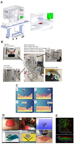
Skin bioprinter prototype. (A) Schematic demonstrating scale, design, and components of the skin bioprinter. (B) The main components of the system consist of 260 µm diameter nozzles, driven by up to eight independently dispensing systems connected to a print-head with an XYZ movement system, in addition to the 3D wound scanner. All components are mounted on a frame small enough to be mobile in the operating room. (C) Skin bioprinting concept. Wounds are first scanned to obtain precise information on wound topography, which then guides the print heads to deposit specified materials and cell types in appropriate locations (Images courtesy of LabTV - National Defense Education Program, Washington, D.C.). (D) Example of skin bioprinting process, where markers that are placed around the wound area used as reference points: (a) prior to scanning with a hand-held ZScanner Z700 scanner (b). Geometric information obtained via scanning is then inputted in the form of an STL file to orient the scanned images to a standard coordinate system (c). The scanned data with its coordinate system is used to generate the fill volume and the path points for nozzle head to travel to print the fill volume (d). Output code is then provided to the custom bioprinter control interface for the generation of nozzle path needed to print fill volume (e, f). (E) This system facilitates the depositing of multiple cell types with high precision and control. The layering of fibroblasts (green) and keratinocytes (red) is shown.
The new bioprinter from WFIRM combines the delivery systems of inkjet printers with mobility and the scanning capability of different topographies. The research team outlined its design and printing approach in the paper “Bioprinting of Autologous skin Cells Accelerates Wound Healing of extensive excisional Full-thickness Wounds.” The main components of the system are a handheld 3D scanner and a print head that can move in any XYZ direction. The print head contains eight 260 μm diameter nozzles, each driven by an independent dispensing motor. The printer components are mounted on a small frame which can be used in an operating room. The system is 79 cm wide (patient head-to-toe direction) by 77 cm deep (cross-patient direction). The system includes a robotic arm and, when fully extended, adds an additional 50 cm—combining for a full reach of 127 cm.
The scanner attached to the printer system is the ZScanner Z700 scanner from 3DSystems. As noted, the scanner is handheld and easy to use. It can capture the entire wound in one continuous scan which results in a computer model of the wound. The model is processed via Geomagic Studio and imported into Artcam 3D software to obtain the full volume and the nozzle path needed to print the fill volume. The wound is split into Z axis layers for its depth, corresponding to dermis and epidermis layers, and each Z-axis layers is overlaid with XY lines that cover the entire wound.
The cartridge-based delivery system is like that used in traditional inkjet printing. The pressure-based delivery system uses three components to print on the wound.
Delivery Mechanism
- The delivery system is based on cartridges that can contain any material to be delivered via the print-head nozzles.
Material Reservoirs
- Each nozzle is connected to a separate cartridge, and each cartridge contains a matrix of fibrinogen and collagen. Separate atomizing nozzles deposit thrombin on the fibrinogen matrix to produce fibrin simultaneously.
Pressure Source
- Air from the pressure source fills the reservoir and drives the material to the delivery mechanism. The system is pressurized with 6.89 kPa of compressed air using a two-stage control valve. Individual syringe pumps are used to achieve identical piston-driven pressure delivery, but with more control over tamping and depressurization.
Based off this delivery system, the keratinocytes and fibroblasts are mixed into a hydrogel and delivered directly on top of the wound layer by layer, replicating the skin structure and accelerating the healing of normal skin and function. The research team has had successful results on mice in clinical trials. The new skin begins to form outward from the center of the wound and, by using the subject’s own unaffected cells for new cell delivery, it allowed the subject’s body to accept the new skin and prevent tissue rejection.
“The technology has the potential to eliminate the need for painful skin grafts that cause further disfigurement for patients suffering from large wounds or burns,” said WFIRM Director Anthony Atala, M.D., a co-author of the paper.
“If you deliver the patient’s own cells, they do actively contribute to wound healing by organizing up front to start the healing process much faster,” added James Yoo, M.D., Ph.D., who led the research team and co-authored the paper with Atala. “While there are other types of wound healing products available to treat wounds and help them close, those products do not actually contribute directly to the creation of skin.”
The next step is to conduct a clinical trial on humans, and hopefully replace the traditional donor or artificial skin graft treatment for the patient’s own 3D-printed skin.

