Diaper Compound Boosts Power of Microscopes
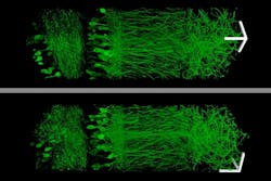
Researchers at the Massachusetts Institute of Technology recently discovered that adding a modified form of the superabsorbent chemical used in disposable diapers can expand samples of brain tissues to 4.5 times their original size. This process, called expansion microscopy, could let doctors take high-resolution images of healthy and diseased tissue using common microscopes.
In the discovery, scientists linked brain-cell proteins to a mesh of sodium Polyacrylate (the diaper absorbent) and added water. The polyacrylate mesh swelled, greatly expanding the size of protein complexes while preserving their normal structural arrangement in the cell. This let the scientists see previously hidden submicroscopic details of cell structures.
According to the National Institute of Health’s acting director of Neurological Disorders and Stroke, Walter Koroshetz, “This kind of outside-the-box technical innovation expands the capability of microscopes widely used in the scientific community to explore the fine structure of the nervous system in health and disease.”
The researchers also developed a special labeling molecule that let them visualize protein complexes and link them to superabsorbent meshes. They also tried out their new visualization method on rodent brain slices and brain cells grown in petri dishes. In each case, the scientists fixed cell proteins in place with formaldehyde and then stripped the cells of their fatty membranes before treating them with labeling molecules. Before treating with acrylate, the scientists took pictures of certain parts of the cells with a high-powered microscope designed to capture fine protein complex details. Then they applied the acrylate, polymerized it to form a mesh, used enzymes to clear away unlinked proteins from the mesh, and expanded the structures with water. After expansion, they took pictures of the same locations using a lower-powered microscope. Comparisons showed that expansion microscopy improved the second microscope’s ability to resolve details of the cells’ proteins.
